Ocular Health/Emergency Eye Exams
WHY ARE REGULAR EYE EXAMS IMPORTANT?
Early eye disease diagnosis and management is essential to preventing irreversible vision loss. Most ocular diseases progress gradually and can be difficult to detect any initial vision changes, but at the same time, they can cause irreversible vision loss if left undiagnosed for too long. In fact, many people, do not realize the loss of vision until the damage is substantial and irreversibly where treatment options may be limited and challenging, and potentially, less effective.
Regular exam with your optometrist allows for close monitoring for ocular health issues and abnormalities. Detection of eye diseases, providing treatment options, and professional management before you may notice any changes can prevent irreversible vision loss and blindness.
Don’t remember when your last exam was? Then it’s probably time to book an appointment.
OPTOMAP – 3D RETINAL PHOTOGRAPH
With this new technology, we are able to photograph over 80% of the retina with a single photo. This allows us to educate you on your specific findings with a detailed explanation and an interactive picture taken during pretesting. This will provide a baseline for how your eye changes, or as a way to monitor abnormal findings over time. This technology is above and beyond the standard of care for comprehensive eye exams so not all clinics are required to have it. At Louie Eyecare we pride ourselves on keeping up with current technology to provide the utmost care. If you haven’t had a comprehensive eye exam in some time, or have never before been shown what the inside of your own eye looks like, we welcome you to come in and be amazed.
OCT (OPTICAL COHERENCE TOMOGRAPHY)
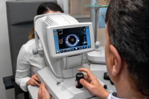
OCT is a non-invasive image that uses light waves to take cross-section pictures of your retina and obtain a clear picture of the structure of your retina and optic nerve, allowing us to check for any signs of diseases like age-related macular degeneration and glaucoma. With it, we can measure and map individual layers of cells in the retina in microns. This greatly aids in the measuring, monitoring, diagnosis, and treatment of inner eye conditions like glaucoma, macular degeneration, and diabetic eye disease. Our Carl Zeiss OCT has allowed us to keep patients in the office for treatment and diagnosis instead of needing a referral to a specialist.
VISUAL FIELD
Our Zeiss Humphrey Visual Field Analyser is the gold standard for detecting visual field deficiencies in diseases like Glaucoma, Macular Degeneration, Optic Neuritis, and damage to the visual cortex of the brain. This test can be very helpful to obtain over time to assess for any changes in visual function.
There are a few things that you can do to prepare for an eye exam. Come with a list of all the medications you are currently taking and any information about your prescription for your current glasses or contact lenses.
Age: All Ages
- Optomap 3D Retinal Photograph: This type of retinal imaging technology is non-invasive and painless. It provides a wide-field, high-resolution view of the back of the eye, including the retina and optic nerve. This technology captures a digital retina image, allowing us to examine and analyze the eye’s health in greater detail.
- Visual acuity test: This test measures your ability to see letters or symbols on a chart from a specific distance.
- Refraction test: This test determines your eyeglass or contact lens prescription by measuring how light bends as it enters your eyes.
- Eye muscle movement test: This test checks your eyes’ alignment and how well they move together.
- Pupil dilation: This test involves using eye drops to dilate your pupils, allowing the eye doctor to examine the retina and optic nerve. This is also sometimes also done in children for the purpose of determining the full prescription amount. This will be done if deemed necessary by the doctor during the exam.
- Tonometry: This test measures the pressure inside your eyes, which can help detect glaucoma.
- Eye health evaluation: The eye doctor will examine your eyes’ external and internal structures to look for any signs of disease or abnormality.
- Assessment and Plan: your results will be explained to you and a plan provided to help out with your specific vision or ocular health findings.
Price: $135
West Edmonton Vision Clinic
Visit our vision clinic in central West Edmonton for comprehensive eye exams, contact lens fittings, glasses, and more. Louie Eyecare Centre is dedicated to providing the highest quality optometric services and products to our patients. Our team of experienced optometrists is here to help you with all of your eye care needs. Schedule an appointment today!
Clinic Hours
Monday Closed
Tuesday 9:00-5:00
Wednesday 9:00-5:00
Thursday 9:00-5:00
Friday 9:00-5:00
Saturday 9:00-2:00
Closed Sunday / Holidays
OUR CLIENTS' FEEDBACK
Frequently Asked Questions
Wearing glasses or contacts can indeed affect dry eye symptoms, but the impact varies. Glasses can help shield the eyes from environmental factors that exacerbate dry eye, such as wind or air conditioning. On the other hand, contact lenses can sometimes worsen dry eye symptoms by absorbing tear moisture or by causing irritation. Certain types of contact lenses are designed to be more breathable and retain moisture better, which may be suitable for people with dry eyes. It’s crucial to discuss with an eye care professional to find the most appropriate type of contact lens or glasses. Proper care and hygiene when using contacts, along with regular breaks from screen use, can help minimize dry eye symptoms.
Dry eye syndrome can be both a temporary condition and a chronic disease, depending on its cause and severity. Environmental factors or certain life situations, such as screen use or air travel can cause temporary dry eye. Chronic dry eye, on the other hand, may result from systemic diseases, medication side effects, or age-related changes in tear production. Management and treatment can alleviate symptoms, but chronic dry eye often requires ongoing therapy. It’s important to consult with an eye care professional for an accurate diagnosis and treatment plan. Understanding the underlying cause is key to determining whether dry eye syndrome will be a temporary issue or a chronic condition.
Yes, some specific exercises and therapies can help relieve dry eye symptoms. Blinking exercises, for example, can help improve meibomian gland function and tear film stability. Warm compresses applied to the eyes can also stimulate tear production and release oils from the glands in the eyelids. Gentle eyelid massages can help spread the oils evenly across the eye surface, reducing dryness. Using a humidifier to add moisture to the air and taking regular breaks to rest the eyes during screen time can also be beneficial. Newer technologies such as IPL (Intense Pulsed Light) and RF (Radio Frequency) are also becoming available. Consulting with an eye care professional for personalized advice on exercises and therapies is recommended.
Sleep plays a crucial role in managing dry eye syndrome. Poor sleep can lead to insufficient eye lubrication and worsening dry eye symptoms. During sleep, the eyes rejuvenate and produce the moisture needed for the next day. Good sleep hygiene practices can help ensure the eyes are well-rested and hydrated. It’s also important to avoid sleeping with any airflow directly hitting the face, as this can dry out the eyes. Establishing a regular, restful sleep schedule can significantly improve dry eye symptoms.
Indeed, some medications can exacerbate dry eye symptoms. Diuretics, antihistamines, antidepressants, and some blood pressure medications are known to reduce tear production or alter tear composition. It’s important to review any current medications with a healthcare provider to determine if they could be contributing to dry eye symptoms. Sometimes, alternative medications with fewer dry eye side effects can be prescribed. Always consult with a healthcare professional before making changes to medication regimens. Patients should also stay hydrated and consider using artificial tears if taking medications known to cause dryness.
Yes, it is quite common for dry eye symptoms to worsen in certain weather conditions. Dry, windy, or smoky environments can lead to increased tear evaporation, exacerbating symptoms. Conversely, high humidity can sometimes alleviate dry eye symptoms because the air is more saturated with moisture. Cold weather, especially during winter when indoor heaters are used, can also dry out the eyes. It’s advisable to use humidifiers in such conditions to maintain indoor humidity levels. Wearing wraparound glasses or protective eyewear outdoors can help shield eyes from harsh conditions.


